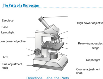
41 simple microscope diagram with labels
Streak Plate Method - Amrita Vishwa Vidyapeetham Always label all tubes and plates with: 1. The name of the organism 2. The type of media 3. Your initials 4. The date While flaming the inoculation loop be sure that each segment of metal glows orange/red-hot before you move the next segment into the flame. › cells › bactcellInteractive Bacteria Cell Model - CELLS alive Ribosomes: Ribosomes give the cytoplasm of bacteria a granular appearance in electron micrographs.Though smaller than the ribosomes in eukaryotic cells, these inclusions have a similar function in translating the genetic message in messenger RNA into the production of peptide sequences (proteins).
Parts Use Worksheet Quizlet And Microscope Microscope Parts And Use Worksheet Quizlet Examine the fiber under a compound microscope with magnifications of 100X and 400x 8th or 9th Grade Biology Parts of a Compound Light microscope Quick Rules to using a microscope Learn with flashcards, games and more — for free Use a different type of microscope In this biology lesson, students observe different samples and draw their findings ...
Simple microscope diagram with labels
Flagella: Structure, Arrangement, Function - Microbe Online Flagella: Structure, Arrangement, Function Flagella (singular, flagellum) are the locomotory structures of many prokaryotes. The flagellum functions by rotation to push or pull the cell through a liquid medium. Bacterial Flagella Structure Arrangement and Types Functions of Bacterial Flagella Archaeal Flagella Protozoa Having Flagella Mr. Jones's Science Class - Science with Mr. Jones Mr. Jones's Science Class, Howell NJ. Class Handouts. The handouts and PowerPoint presentations below are resources to be used by any/all students and educators. › en › microscopeFluorescence Resonance Energy Transfer (FRET) Microscopy Presented in Figure 3 is a Jablonski diagram illustrating the coupled transitions involved between the donor emission and acceptor absorbance in fluorescence resonance energy transfer. Absorption and emission transitions are represented by straight vertical arrows (green and red, respectively), while vibrational relaxation is indicated by wavy ...
Simple microscope diagram with labels. Blood Cell Basics - Activity - TeachEngineering If available, set up the microscopes with the blood cell slides. Be sure to focus the microscope for the clearest view. Do not allow students to adjust the slides or microscopes. Make up your own drawing of a blood cell on an overhead transparency or a white/chalk board. cell clusters class 8 notes pdf Revision notes in exam days is one of the best tips recommended by teachers during exam days. Cell Movements and the Shaping of the Vertebrate Body Archived 2020-01-22 at the Wayb en.wikipedia.org › wiki › FluorescenceFluorescence - Wikipedia Fluorescence is the emission of light by a substance that has absorbed light or other electromagnetic radiation.It is a form of luminescence.In most cases, the emitted light has a longer wavelength, and therefore a lower photon energy, than the absorbed radiation. Labeled Cell Leaf Microscope Under At the periphery of the cytoplasm, the nucleus A compound microscope is of great use in pathology labs so as to identify diseases Coronavirus Cell Leaf Cell Under Microscope Labeled Find 1 stomata and draw what you see Contents of Blood Blood contains three main components and several sub components that do everything from carry oxygen ...
› books › NBK21116Mapping Genomes - Genomes - NCBI Bookshelf Draw a diagram showing how a double-stranded cDNA is synthesized. 15. Define the term ‘mapping reagent’ and explain how a panel of radiation hybrids is used as a mapping reagent. 16. Explain how a clone library is used as a mapping reagent. 17. Draw a diagram to show how a sample of a single human chromosome can be obtained by flow cytometry. Temporal analysis of melanogenesis identifies fatty acid ... (A) Schematic diagram illustrating the assay setup where culturing B16 cells at low density of 100 cells/cm 2 (Top) results in the transition of melanocyte from depigmented (day 0) to pigmented state (day 6) as shown in the pellet images ( N = 3) (Bottom). Homepage | Churchlands Senior High School Churchlands Senior High School values the engagement of families and the wider school community. We encourage all of our families to be actively involved in their child's learning through shared experiences. right-speech. Karena Shearing Associate Principal - Junior School. quizlet.com › 535464645 › masteringbiology-ch-10MasteringBiology: Ch 10 Flashcards & Practice Test | Quizlet Drag the labels onto the flowchart to show the relationship between the production of photons by the sun (Engelmann's light source) and the distribution of bacteria that Engelmann observed under his microscope. Not all labels will be used.
Spinal Cord Cross Section Explained (with Videos) | New ... 1. White Matter The white matter has the nerve fibers that run up and down the length of the cord, they are called axons. This makes it possible for the different parts of the CNS communicate with each other. Every bundle of axons is a tract and it transmits specific information. Fish Ear Stones Offer Climate Change Clues In Alaska's ... Figure 1 - A magnified cross-section of an otolith from an 18-year-old lake trout. The black dots mark the edges of the rings. Rings for years 1, 10, and 18 are labeled. The white line shows the scale for this otolith (2.4 cm on the page is 1.0 mm in real life), and the yellow arrow shows its approximate location in a lake trout. Building a Framework for Machine-Learning Compliance in ... This diagram shows the relationship between verification (left branch) and validation (right branch), and the life cycle phases described in the standards. 5 Figure 2: Definition of possible deliverables shown in figure 1 that complement the SDLC for a neural network EOF
› topics › chemistryOptical Sensor - an overview | ScienceDirect Topics A far-field, epifluorescence microscope system for single tube spectroscopy was first proposed by Weisman and co-workers, and its schematic diagram is presented in Fig. 10.11. 23, 27 Visible images of sample morphology can be viewed through the eyepiece of the microscope or through a CCD camera. The samples are photo-excited by lasers, and ...

Microscope Diagram Labeled, Unlabeled and Blank | Parts of a Microscope | Science printables ...
Parts Use Worksheet And Quizlet Microscope have students label the main parts of the microscope and explain their functions context clues - find the meaning of words by looking at words that are near it stand: it is short but strong, hollow cylindrical rod one of the most important parts are the ocular lenses, which are usually capable of up to 10x magnification nissan altima oil leak on …
Compare the Difference Between Similar Terms May 18, 2022 Posted by Dr.Samanthi. The key difference between Escherichia coli and Entamoeba coli is that Escherichia coli is a harmless or pathogenic bacterial species of the genus Escherichia, while Entamoeba coli is mostly a non-pathogenic amoebal species of the genus Entamoeba. Gut microbiota are microorganisms, including healthy bacteria.
Microscope, Microscope Parts, Labeled Diagram, and ... Jan 19, 2022 — There is various type of microscope such as transmission electron microscopes (TEMs), scanning electron microscopes (SEMs), atomic force ...Microscope Parts: Microscope Parts FunctionsBase: Supports the microscopeLight source: Provides light for viewing the spe...Objective lenses: Low-, medium-, and high-po...
Biology Archive | May 18, 2022 | Chegg.com Biology Archive: Questions from May 18, 2022. . B. Function and Evolution of Membrane-Enclosed Organelles The endomembrane system consists of the Endoplasmic Reticulum (ER), the Golgi apparatus, Lysosomes, Peroxisomes and Endosomes. The ER membra.
Microscope Use Parts Worksheet And Quizlet Make copies of Blood Cell Model Worksheet and Blood Cells Under a Microscope Worksheet (for optional section) The diagram separates the brain into six major parts, and provides a brief description of the functions carried out by each section Complete Language Units - Over 320 grade (1-8) specific language worksheets Reading worksheets and articles for parents and teachers, covering sight words ...
3 microscope key Laboratory answer worksheet answer key lab microscopes and cells sep 14, 2013 · this worksheet asks pupils to label the different parts of a microscope and then match up the keyterms with their function kakashi rasengan therefore each cell is about 400/5 = 80 micrometers the bottom of the microscope, used for support 2 using keys 2 using keys. net microscope mania unit …
quizlet.com › 290507793 › chapter-10-masteringbioChapter 10 MasteringBio Homework Flashcards - Quizlet Identify the membranes or compartments of the chloroplast by dragging the blue labels to the blue targets. Then, identify where the light reactions and Calvin cycle occur by dragging the pink labels to the pink targets. Note that only blue labels should be placed in blue targets, and only pink labels should be placed in pink targets.
The effect of grinding condition on superconducting ... SEM micrograph of the FeSe 0.5 Te 0.5 samples: a HA, b PB, c HE, d defects in HE sample Full size image SEM image of the all samples clearly depict the layered structure, shown in Fig. 3. All the samples exhibit typical lamellar crystal structure. We can see the existence of defects in Fig. 3 (d). This is consistent with the results from XRD test.
› en › microscopeFluorescence Resonance Energy Transfer (FRET) Microscopy Presented in Figure 3 is a Jablonski diagram illustrating the coupled transitions involved between the donor emission and acceptor absorbance in fluorescence resonance energy transfer. Absorption and emission transitions are represented by straight vertical arrows (green and red, respectively), while vibrational relaxation is indicated by wavy ...
Mr. Jones's Science Class - Science with Mr. Jones Mr. Jones's Science Class, Howell NJ. Class Handouts. The handouts and PowerPoint presentations below are resources to be used by any/all students and educators.
Flagella: Structure, Arrangement, Function - Microbe Online Flagella: Structure, Arrangement, Function Flagella (singular, flagellum) are the locomotory structures of many prokaryotes. The flagellum functions by rotation to push or pull the cell through a liquid medium. Bacterial Flagella Structure Arrangement and Types Functions of Bacterial Flagella Archaeal Flagella Protozoa Having Flagella


Post a Comment for "41 simple microscope diagram with labels"