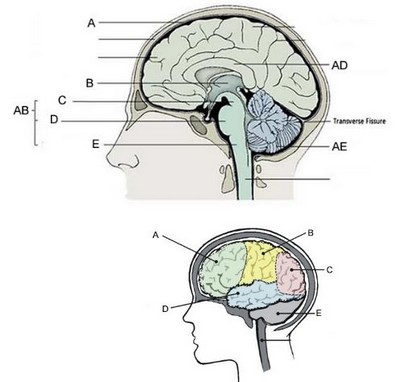
44 the brain with labels
Nervous System - Label the Brain This brain part controls thinking. This brain part controls balance, movement, and coordination. This brain part controls involuntary actions such as breathing, heartbeats, and digestion. This … myclass.theinspiredinstructor.com › science › healthNervous System - Label the Brain This brain part controls thinking. This brain part controls balance, movement, and coordination. This brain part controls involuntary actions such as breathing, heartbeats, and digestion. This part of the nervous system moves messages between the brain and the body. This part of the cerebrum interprets and sorts information from the senses.
Amazon.com: XINDAM 3D Human Brain with Labels Anatomical Model ... XINDAM 3D Human Brain with Labels Anatomical Model Paperweight(Laser Etched) in Crystal Glass Ball Science Gift (Included LED Base) Brand: XINDAM. 4.8 out of 5 stars 20 ratings. $66.99 $ 66. 99 & FREE Returns . Return this item for free. Free returns are available for the shipping address you chose. You can return the item for any reason in new ...

The brain with labels
Anatomy of the Brain | Simply Psychology The occipital lobes are located at the back of the brain behind the temporal and parietal lobes and below the occipital bone of the skull (Figure 7).. The occipital lobes receive sensory information from the retinas of the eyes which is then encoded into different visual data. Some of the functions of the occipital lobes include being able to assess size, depth, and distance, determine colour ... Parts of the brain: Learn with diagrams and quizzes | Kenhub Labeled brain diagram. First up, have a look at the labeled brain structures on the image below. Try to memorize the name and location of each structure, then proceed to test yourself with the blank brain diagram provided below. Labeled diagram showing the main parts of the brain. Lobes of the brain - Queensland Brain Institute The brain's cerebral cortex is the outermost layer that gives the brain its characteristic wrinkly appearance. The cerebral cortex is divided lengthways into two cerebral hemispheres connected by the corpus callosum. Traditionally, each of the hemispheres has been divided into four lobes: frontal, parietal, temporal and occipital . Although ...
The brain with labels. Diagram Of Brain with their Labelings and Detailed … The midbrain is also called as Mesencephalon. The midbrain is the smallest region of the brain, found at the centre of the brain, between cerebral cortex and hindbrain. It comprises tectum, … Label The Brain - Mr. Barth's Class Label The Brain. The following websites are to help you learn and remember the parts of the brain and their locations. Please go through each of websites and become familiar with each of the parts of the brain. I would advise you to repeat each of them a few times until you have the locations memorized. Click on the link to the left to review ... › nervous › brain_labelBrain Label - The Biology Corner Answers: A = parietal labe | B = gyrus of the cerebrum | C = corpus callosum | D = frontal lobe. E = thalamus | F = hypothalamus | G = pituitary gland | H = midbrain. J = pons | K = medulla oblongata | L = cerebellum | M = transverse fissure | N = occipital lobe. Labels on the Brain – Cognitioneducation Jul 07, 2020 · This is your brain, on labels. To explain your brain on labels, I’ll start small, really small. Our brains are made of neurons, billions of them, and our experiences are represented …
Positions and Functions of the Four Brain Lobes - MD-Health.com The occipital lobe, the smallest of the four lobes of the brain, is located near the posterior region of the cerebral cortex, near the back of the skull. The occipital lobe is the primary visual processing center of the brain. Here are some other functions of the occipital lobe: Visual-spatial processing. Movement and color recognition. Parts of the brain: Learn with diagrams and quizzes Oct 28, 2021 · Labeled brain diagram. First up, have a look at the labeled brain structures on the image below. Try to memorize the name and location of each structure, then proceed to … Brain Label (Remote) - The Biology Corner Brain Label (Remote) Shannan Muskopf December 29, 2020. This brain labeling activity was created for remote learners as an alternative to the labeling and coloring worksheet we would traditionally do in class. Instead of coloring and labeling on printouts, students use google slides to drag labels to the images or type the answers into text boxes. Ventricles of the Brain: Labeled Anatomy, Function, CSF Flow ... Learn the ventricles of the brain along with their definition, function, location, anatomy, and cerebrospinal fluid (CSF) flow using labeled diagrams. The ventricular system contains the lateral, third, and fourth ventricles whose function is to produce cerebrospinal fluid. Learn where CSF is found,
3D Brain This interactive brain model is powered by the Wellcome Trust and developed by Matt Wimsatt and Jack Simpson; reviewed by John Morrison, Patrick Hof, and Edward Lein. Structure descriptions were written by Levi Gadye and Alexis Wnuk and Jane Roskams . Brain Label | Human anatomy and physiology, Ap biology, Basic anatomy ... This shows a kidney and the nephron with arrows that point to specific structures within the kidney for students of anatomy to label. Image of the ankle and wrist showing the tarsals and the carpals; students label the bones. Notes over the respiratory system with powerpoint presentation and images for labeling. Applications of Arterial Spin Labeled MRI in the Brain - PMC Because the T1 relaxation rate for water in blood or tissues is on the order of 1-2 seconds, only small amounts of arterial spin labeled water accumulate in the brain, and prolongation of T1 with field strength represents a major benefit of high-field MRI for ASL studies. Fortunately, 3 Tesla MRI machines are now widespread. Labeled Diagrams of the Human Brain You'll Want to Copy Now The height of the human brain is about 3.6 inches and it weighs about 4 to 5 lbs at birth and 3 lbs in adults. The total surface area of the cerebral cortex is about 2,500 cm2 and when stretched, it will cover the area of a night table. The brain is composed of 77 to 78% water and 10 to 12% lipids. It contains 8% proteins 1% carbohydrates, 2% ...

35 Label The Brain Diagram - Labels Design Ideas 2020
Label the Brain Anatomy Diagram Flashcards | Quizlet Start studying Label the Brain Anatomy Diagram. Learn vocabulary, terms, and more with flashcards, games, and other study tools. Home. Subjects. Explanations. Create. ... the part of the brain-stem that joins the hemispheres of the cerebellum and connects the cerebrum with the cerebellum; regulates sleep and dreams.
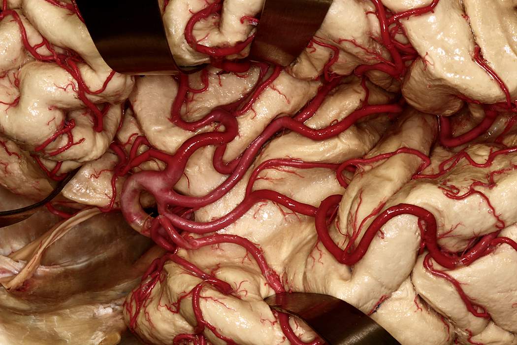
Lateral Exposure of Middle Cerebral Artery in Sylvian Fissure | Neuroanatomy | The Neurosurgical ...
Frontiers | 101 Labeled Brain Images and a Consistent Human Cortical ... We introduce the Mindboggle-101 dataset, the largest and most complete set of free, publicly accessible, manually labeled human brain images. To manually label the macroscopic anatomy in magnetic resonance images of 101 healthy participants, we created a new cortical labeling protocol that relies on robust anatomical landmarks and minimal manual edits after initialization with automated labels ...
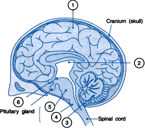
Label parts 1 to 6 in the given figure of the brain. - Zigya
› anatomy-of-the-brainBrain Anatomy and How the Brain Works - Hopkins Medicine The cerebellum (“little brain”) is a fist-sized portion of the brain located at the back of the head, below the temporal and occipital lobes and above the brainstem. Like the cerebral cortex, it has two hemispheres. The outer portion contains neurons, and the inner area communicates with the cerebral cortex.

brain label and function(dots outside of brain) Quiz
bananasformentera.com › 40-diagram-of-the-brain40+ Diagram Of The Brain With Labels And Functions Pics Apr 30, 2021 · 40+ Diagram Of The Brain With Labels And Functions Pics. It is made up of more than 100 billion nerves that communicate in trillions. To list all the functions and responsibilities of this collection of billions of neurons, you need to compose a whole book. Colored And Labeled Human Brain Diagram Stock Illustration … from media.istockphoto.com

Control of Drosophila Type I and Type II central brain neuroblast proliferation by bantam ...
› brain › picture-of-the-brainBrain (Human Anatomy): Picture, Function, Parts, Conditions ... The brain is one of the largest and most complex organs in the human body. It is made up of more than 100 billion nerves that communicate in trillions of connections called synapses. • The ...
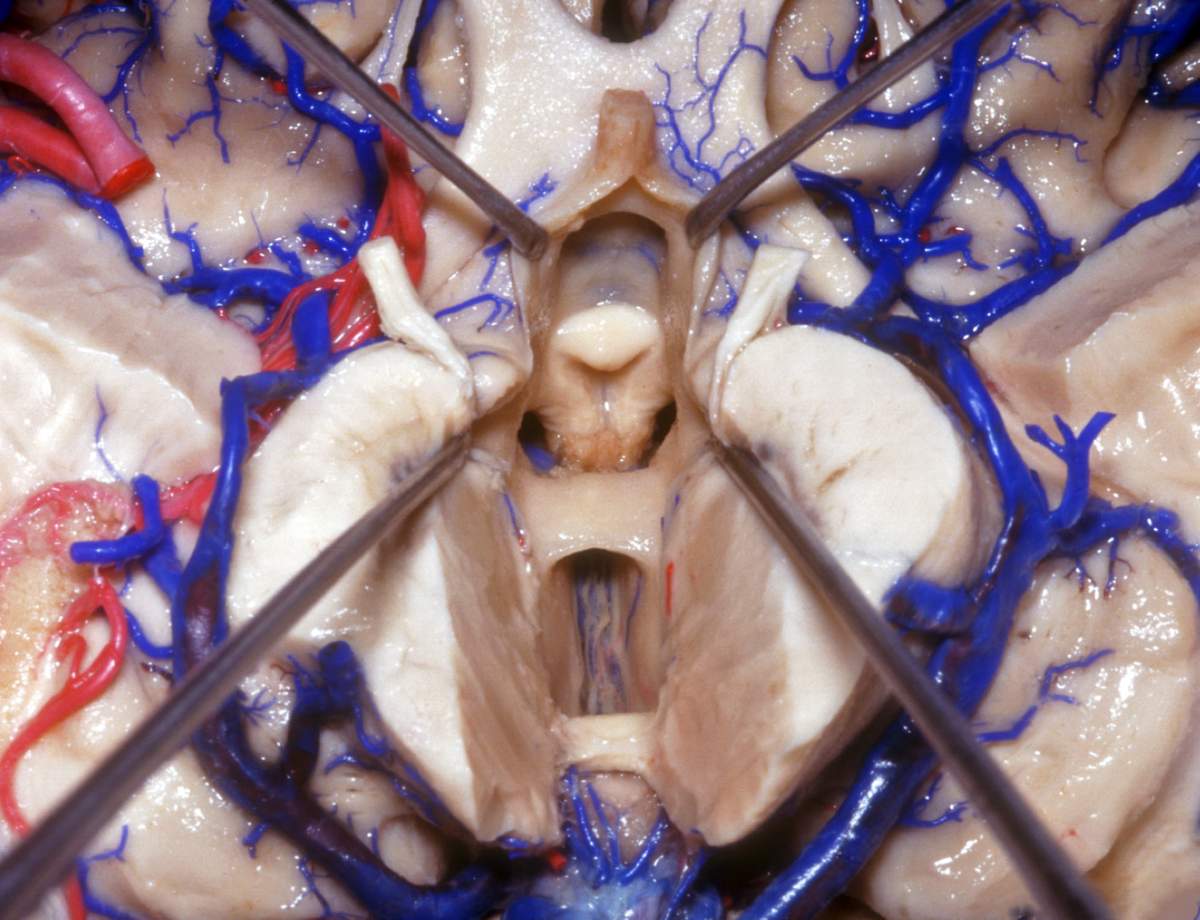
Midline Inferior View of Sectioned Third Ventricular Floor | Neuroanatomy | The Neurosurgical ...
Brain Label (Remote) - The Biology Corner Dec 29, 2020 · Brain Label (Remote) Shannan Muskopf December 29, 2020. This brain labeling activity was created for remote learners as an alternative to the labeling and coloring …

20 Image Quotes To Share / Digital Information World
Brain: Anatomy, Pictures, Functions, and Conditions The Brain Stem. PIXOLOGICSTUDIO/SCIENCE PHOTO LIBRARY / Getty Images. The brainstem is an area located at the base of the brain that contains structures vital for involuntary functions such as the heartbeat and breathing. The brain stem is comprised of the midbrain, pons, and medulla. 3.

Dr Balaji Anvekar FRCR: Diffuse Axonal Injury CT Brain
Labeled Diagrams of the Human Brain You’ll Want to … The height of the human brain is about 3.6 inches and it weighs about 4 to 5 lbs at birth and 3 lbs in adults. The total surface area of the cerebral cortex is about 2,500 cm2 and when stretched, …

Figure 1. Rates of Gray Matter Loss in Normal Adolescents and Matched Subjects with ...
paintingvalley.com › drawing-of-the-brain-with-labelsDrawing Of The Brain With Labels at PaintingValley.com ... Tags: brain, labels All rights to paintings and other images found on PaintingValley.com are owned by their respective owners (authors, artists), and the Administration of the website doesn't bear responsibility for their use.

99 best Brain and Mind images on Pinterest | The brain, Mental health and Neuroscience
Brain-Based Labels Bunk? | The Scientist Magazine® Brain-Based Labels Bunk? An fMRI study shows speculations that people are "left-brained" versus "right-brained" are not backed by evidence. Kate Yandell. Aug 19, 2013. ... "But people don't tend to have a stronger left- or right-sided brain network. It seems to be determined more connection by connection."
Brain Label
Brain Records - Wikipedia Brain was a Hamburg-based record label prominent in the 1970s releasing several important Krautrock records by bands such as Neu!, Cluster and Guru Guru.Many of its more prominent records are currently being reissued on CD by Repertoire Records.. In the middle of 1971, Rolf-Ulrich Kaiser's management style at Ohr caused two of his A&R men, Bruno Wendel and Günter Körber, to leave Ohr and set ...

The Human Brain: Introduction
Labeled Brain Model Diagram | Science Trends Cerebrum. The cerebrum is the largest and most complex portion of the human brain. The cerebrum's function is to control our actions and thoughts, either conscious or unconscious, and responses to stimuli. The cerebrum itself is typically divided into four different lobes: the temporal lobe, the parietal lobe, the occipital lobe, and the ...
Human Brain Anatomy - YouTube
Brain Anatomy and How the Brain Works | Johns Hopkins … The cerebellum (“little brain”) is a fist-sized portion of the brain located at the back of the head, below the temporal and occipital lobes and above the brainstem. Like the cerebral cortex, it …

Simplified Brain Labeled Decal | Shop Fathead Anatomical Images Graphics
Brain - Human Brain Diagrams and Detailed Information The brain needs to store many different types of information that it receives from the senses and that it develops through thinking in the association areas. Information in the brain is stored in a few different ways depending on its source and how long it is needed. Our brain maintains short-term memory to keep track of the tasks in which the ...

Brain WebQuest - GW8science
DOC Label the Brain Anatomy Diagram - Windsor C-1 School District Answers: Label the Brain Diagram The Brain. Read the definitions below, then label the brain anatomy diagram. Cerebellum - the part of the brain below the back of the cerebrum. It regulates balance, posture, movement, and muscle coordination. Corpus Callosum - a large bundle of nerve fibers that connect the left and right cerebral hemispheres.
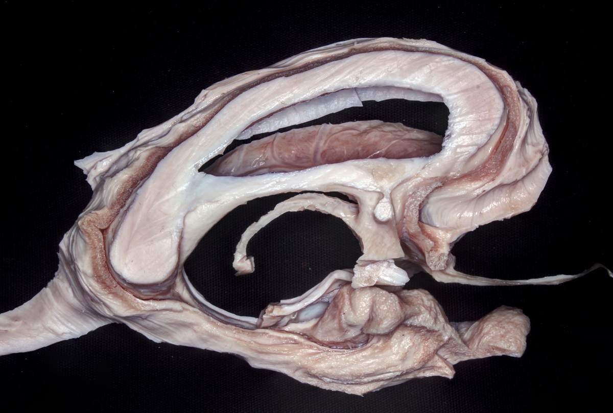
Dissected Corpus Callosum and Fornix | Neuroanatomy | The Neurosurgical Atlas, by Aaron Cohen ...
Lobes of the brain: Structure and function | Kenhub The motor cortex corresponds to the precentral gyrus of the frontal lobe. The precentral gyrus contains the primary motor cortex (Brodmann area 4), which is responsible for integrating signals from different brain regions to modulate motor function. The primary motor cortex is where the corticospinal tract originates. Anterior to the primary motor cortex of the precentral gyrus is the premotor ...

brain labeled
cognitioneducation.me › 2012/02/24 › labels-on-the-brainLabels on the Brain – Cognitioneducation This is your brain, on labels. To explain your brain on labels, I'll start small, really small. Our brains are made of neurons, billions of them, and our experiences are represented in the form of complex inter-connections among neurons. When you have an experience, select networks of neurons in your brain become active, enabling you to ...
Post a Comment for "44 the brain with labels"