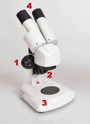
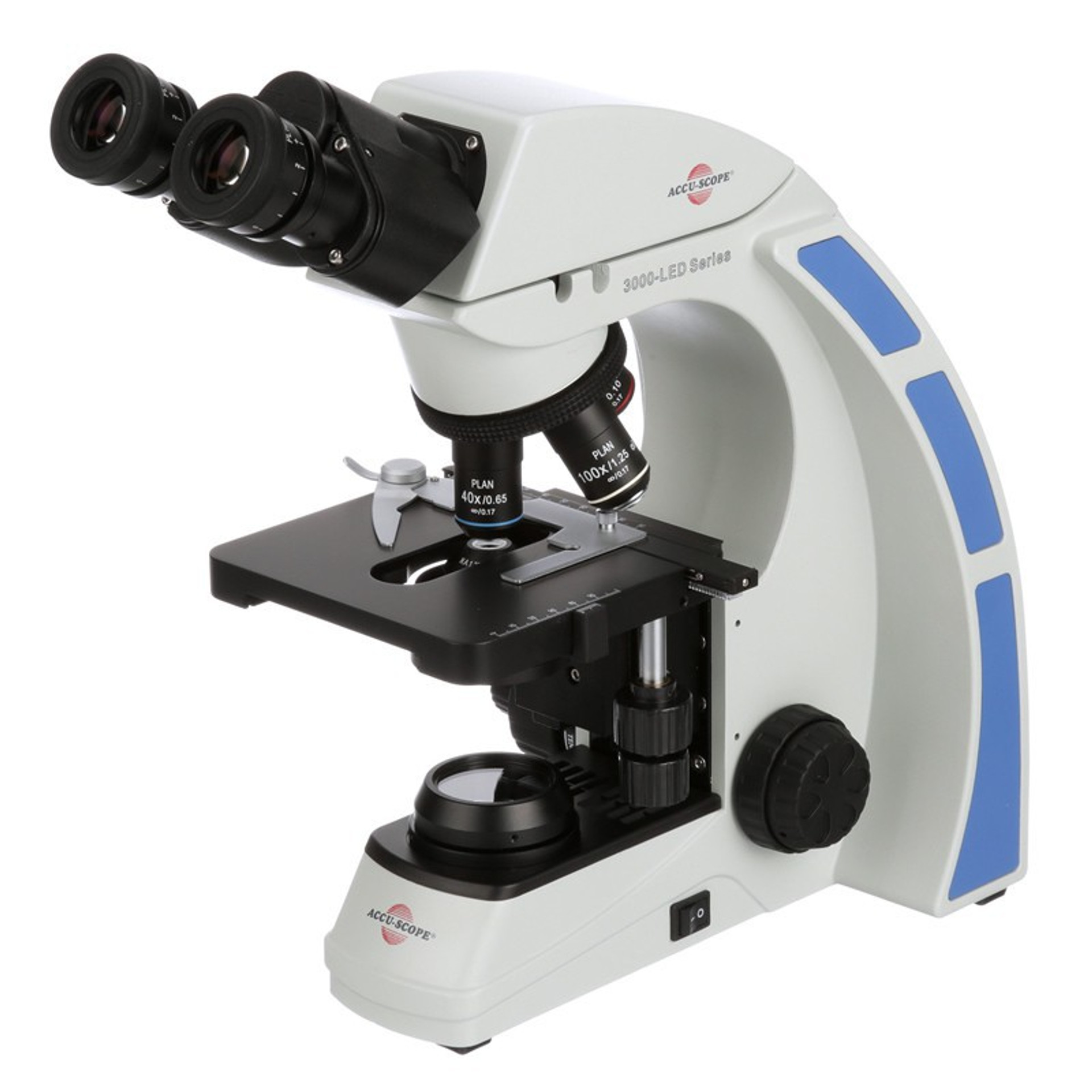
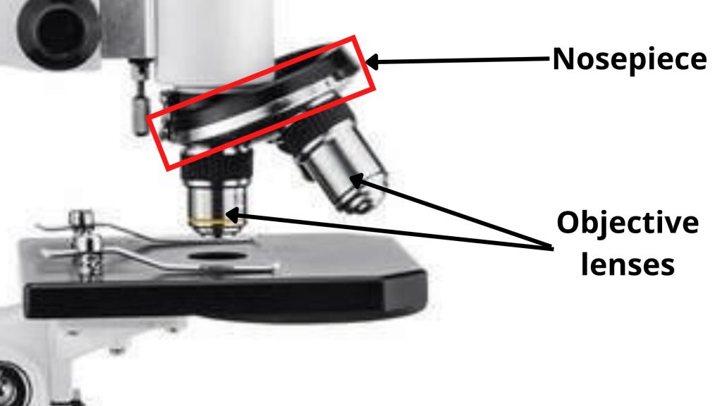
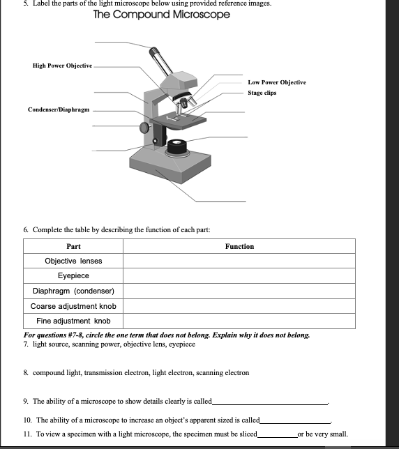
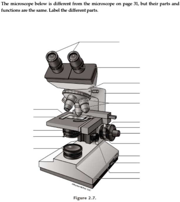
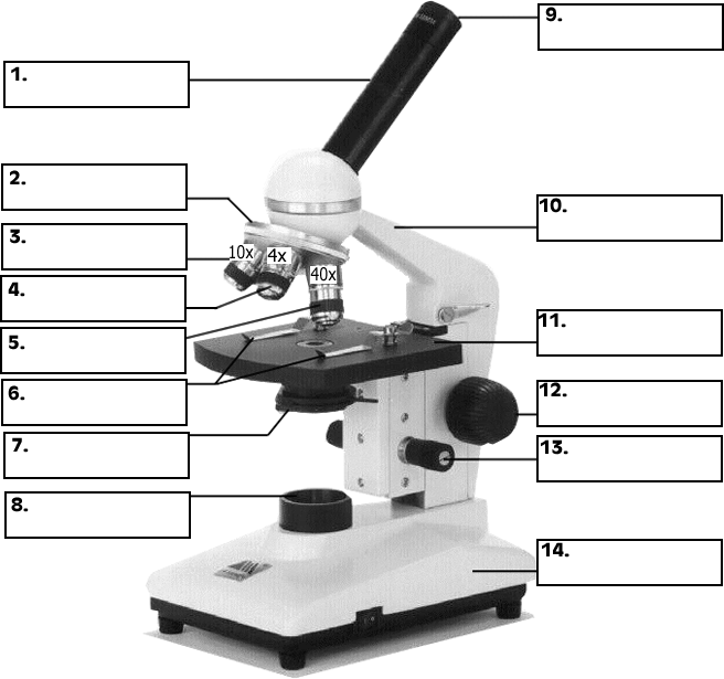
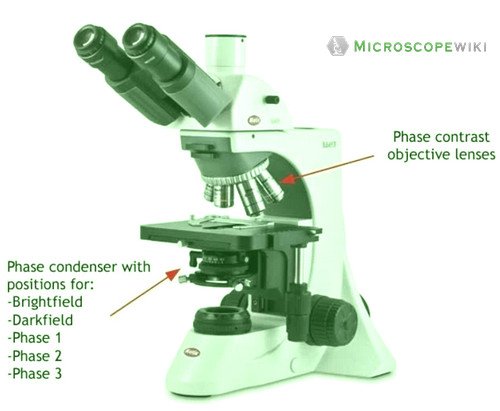
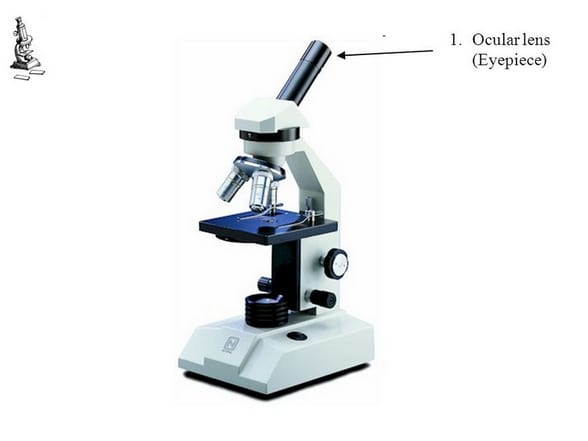
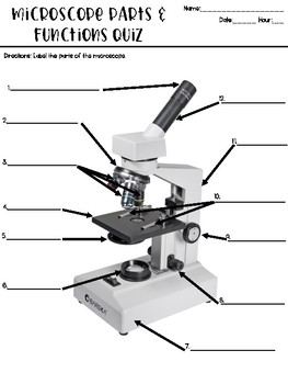
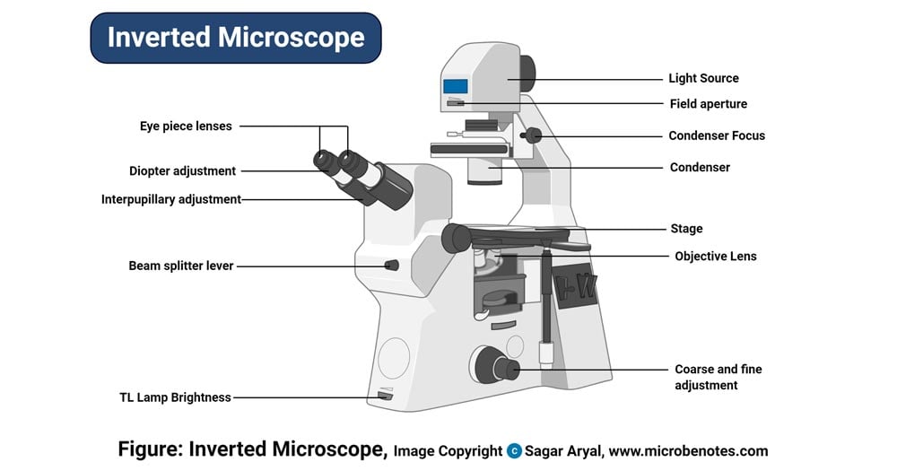
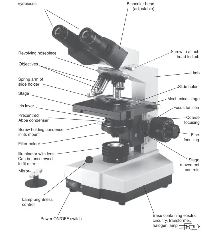
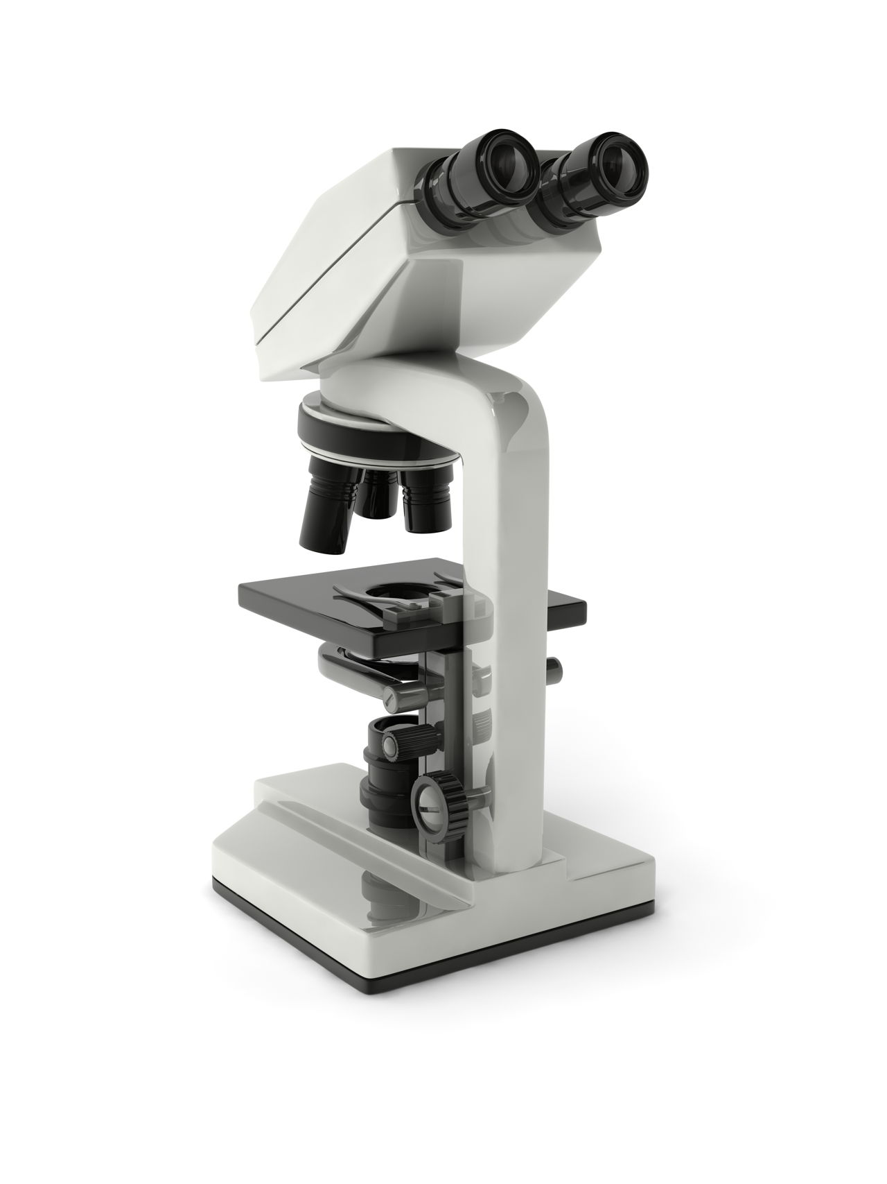
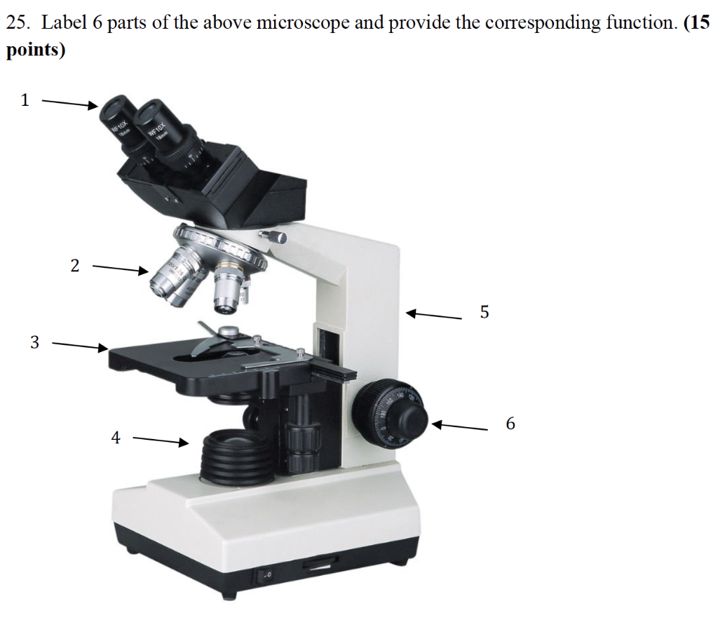
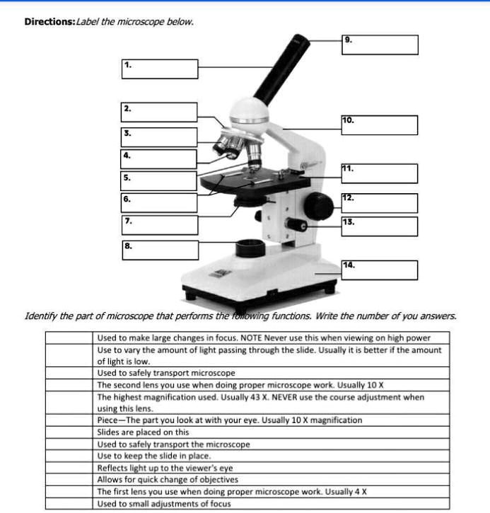
40 microscope with labels and functions
ORDER BY clause - Azure Databricks - Databricks SQL nulls_sort_order. Optionally specifies whether NULL values are returned before/after non-NULL values. If null_sort_order is not specified, then NULLs sort first if sort order is ASC and NULLS sort last if sort order is DESC. NULLS FIRST: NULL values are returned first regardless of the sort order. NULLS LAST: NULL values are returned last ... Autoclave: Sterilization Principle, Procedure, Types, Uses Power Switch: It is present at the side of the autoclave and controls the electricity supplied to the auoclave. Control Panel: It controls the pressure and temperature inside the vessel and is present beside the main switch. Water Level Indicator: It helps indicate the water level of the autoclave. The correct level of water is essential.
cardiac muscle | Definition, Function, & Structure | Britannica Cardiac muscle cells form a highly branched cellular network in the heart. They are connected end to end by intercalated disks and are organized into layers of myocardial tissue that are wrapped around the chambers of the heart. The contraction of individual cardiac muscle cells produces force and shortening in these bands of muscle, with a resultant decrease in the heart chamber size and the ...
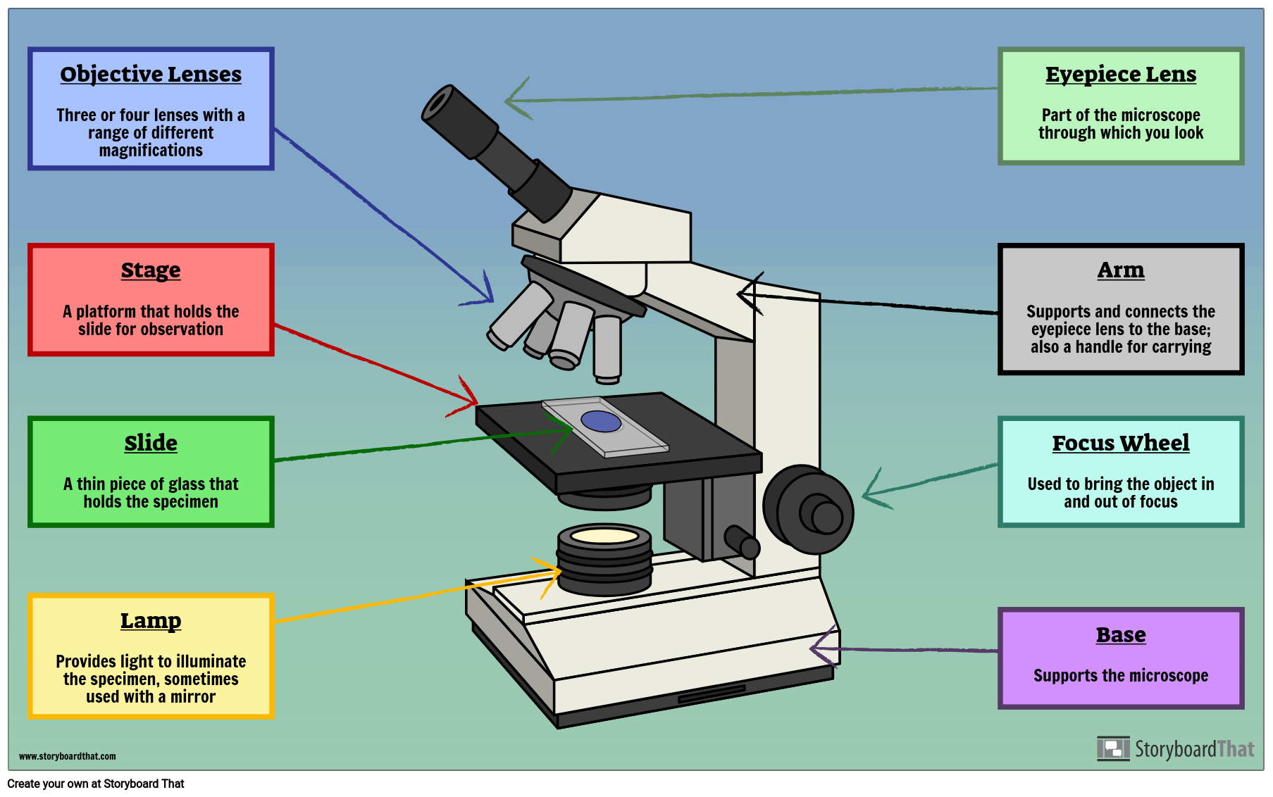
Microscope with labels and functions
Cell biology Virtual Lab II - Amrita Vishwa Vidyapeetham To stain lignin of the plant section and observe under the microscope. Principle . Lignin is a chemical compound derived from wood and is found in the secondary cell walls of plants. It is a polymer of aromatic subunits derived from phenylalanine. Lignin is found in the spaces in the cell wall between cellulose, hemicelluloses and pectin ... Nasal mucosa: structure, function and labeled diagram | GetBodySmart Micrograph of respiratory mucosa: Swipe to show/ hide labels 1 2 Physiology of the Respiratory Mucosa (Nasal Mucosa) As air passes over the nasal mucosa, it is prepared or conditioned to safely pass deeper into the respiratory system. The heat radiated from the blood vessels in the lamina propria warms the air to near body temperature. Microscope Mania Worksheet Answers Key › Athens Mutual Student Corner Use the word list to help you label the microscope. Read the directions for each section on the cards provided at. Microscope quiz with answer key! Worksheet identifying the parts of the compound light microscope answer key 1 body tube 2 revolving nosepiece 3 biology labs microscope parts microscopic.
Microscope with labels and functions. pch in R: How to Use Plot Character in R - R-Lang The pch in R defines the point symbols in the functions plot () and lines (). The pch stands for plot character. The pch contains numeric values ranging from 0 to 25 or character symbols ("+", ".", ";", etc.) specifying in symbols (or shapes). The pch is the standard argument to set the character plotted in several R functions. What features and label should I put in center loss and softmax function? alternates between positive and negative pairs. ''' pairs = [] labels = [] n = min ( [len (digit_indices [d]) for d in range (10)]) - 1 for d in range (10): for i in range (n): z1, z2 = digit_indices [d] [i], digit_indices [d] [i + 1] pairs += [ [x [z1], x [z2]]] inc = random.randrange (1, 10) dn = (d + inc) % 10 z1, z2 = digit_indices … MATHEMATICA TUTORIAL, Part 1.1: Labeling Figures - Brown University There are times when the axes could interfere with displaying certain functions and solutions to ODEs. Fortunately, getting rid of axes in recent versions of Mathematica is very easy. One method of specifying axes is to use the above options, but there is also a visual method of changing axes. Motility Test (Theory) - Amrita Vishwa Vidyapeetham Virtual Lab Fig:-Lophotrichous flagellum seen under light microscope . Flagella are spread fairly evenly over the whole surface of peritrichous bacteria . Fig:-Peritrichous flagellum seen under light microscope . When anticlockwise rotation is resumed, the cell tends to move in a new direction. This ability is important, since it allows bacteria to change ...
Layers of the Stomach | New Health Advisor The main job of the mucosa is to secrete mucus that protects the stomach from its own acids. In this layer, small pores known as gastric pits are responsible for creating the acids that the mucosa protects the stomach from. And the muscularis tissue in it helps the mucosa form folds to further protect the stomach. 2. Submucosa Kidney Structures and Functions Explained (with Picture and Video ... Kidney Function. The urinary system depends on proper kidney structure and function. Some of these core actions include: Excretes waste: The kidneys get rid of toxins, urea, andexcess salts. Urea is a nitrogen-based waste product of cell metabolism that is produced in the liver and transported by the blood to the kidneys. Light Microscope - Amrita Vishwa Vidyapeetham Virtual Lab Optical microscopes function on the basis of optical theory of lenses by which it can magnifies the image obtained by the movement of a wave through the sample. The waves used in optical microscopes are electromagnetic and that in electron microscopes are electron beams. 5 White Blood Cells Types and Their Functions - New Health Advisor Function: Eosinophils work by releasing toxins from their granules to kill pathogens. The main pathogens eosinophils act against are parasites and worms. High eosinophil counts are associated with allergic reactions. 5. Basophils Basophils are the least frequent type of white blood cell, with only 0-100 cells per mm 3 of blood.
5. Preparation Diopter adjustments (1) - Nikon Instruments Inc. Please do the following. ① Using the 10X objective, rotate the focus knobs, and bring the specimen in focus. ② Switch to the 40X objective lens and turn the fine focus knob to refocus on the sample. ③ Switch to the 10X objective again, adjust the focus by individually rotating only the diopter rings on the left and right eyepieces. Boost LabVIEW Productivity with Quick Drop - NI Select a VI, then press to open Quick Drop. Once the window appears press to delete the selected VI and connect corresponding input and output wires. Some LabVIEW programmers prefer their labels on the left of input and right of outputs, and changing those positions can take time. Automate the process by pressing Ultrastructure of cells quiz 1.2 - Subscription websites for IB ... If you found a eukaryote cell in an electron microscope image, and it contained a lot of rER, Golgi apparatus and many darkly stained vesicles, what do you think the function of the cell is most likely to be? A. The production and transmission of a nerve impulse. B. The transport of oxygen in the blood. C. The storage of lipids. D. Microscope Parts and Lenses Trivia Quiz | Miscellaneous Science | 10 ... Quiz Answer Key and Fun Facts 1. What is the light source of a microscope also known as? Answer: Illuminator The illuminator, or light source, is located under the stage of a microscope. It is either made of mirrors or an electric light. The object of the light is to give you a bright background so you can more easily see the object on the slide.
Microscope Mania Worksheet Answers Key › Athens Mutual Student Corner Use the word list to help you label the microscope. Read the directions for each section on the cards provided at. Microscope quiz with answer key! Worksheet identifying the parts of the compound light microscope answer key 1 body tube 2 revolving nosepiece 3 biology labs microscope parts microscopic.
Nasal mucosa: structure, function and labeled diagram | GetBodySmart Micrograph of respiratory mucosa: Swipe to show/ hide labels 1 2 Physiology of the Respiratory Mucosa (Nasal Mucosa) As air passes over the nasal mucosa, it is prepared or conditioned to safely pass deeper into the respiratory system. The heat radiated from the blood vessels in the lamina propria warms the air to near body temperature.
Cell biology Virtual Lab II - Amrita Vishwa Vidyapeetham To stain lignin of the plant section and observe under the microscope. Principle . Lignin is a chemical compound derived from wood and is found in the secondary cell walls of plants. It is a polymer of aromatic subunits derived from phenylalanine. Lignin is found in the spaces in the cell wall between cellulose, hemicelluloses and pectin ...


(159).jpg)











Post a Comment for "40 microscope with labels and functions"